The Department maintains an impressive collection of facilities for Mass Spectrometry, NMR Spectroscopy, Optical Characterization and X-Ray.
Mass Spectrometry Facility
The Department of Chemistry and Biochemistry operates an in-house Mass Spectrometry facility to support research and teaching activities. Mass Spectrometry is a primary tool for structure elucidation, molecular weight determination and molecular formula confirmation for organic and biological materials. This family of techniques measures the masses of organic and biological molecules by creating gas phase ions from the analytes and manipulating those ions in electric and magnetic fields. Chemical structure may be probed by fragmenting molecular ions and mass measuring the resulting product ions.
A wide range of ionization techniques and capabilities are available. Services are available by sample submission to the facility staff or by direct instrument access for trained users on a 24/7 basis. Facility staff is available for operator training as well as consultation on experimental design and data interpretation.
Instrumentation Available
A Bruker Microflex LRF MALDI TOF mass spectrometer is used for the analysis of a broad range of compounds including small molecules, peptides, proteins, nucleic acids, and synthetic polymers. The Microflex LRF has a mass range of up to 20,000 Da in reflectron mode or up to 500,000 in linear mode. Additionally the instrument is capable of mass spectral imaging and post source decay analysis. Software is available for the analysis of polymer data.
A Waters GCT Premier Time-of-Flight MS is equipped with electron ionization (EI), chemical ionization (CI), and field ionization/field desorption (FI/FD) ion sources. EI, CI, and FI techniques are suitable for volatile and semi-volatile organic compounds with sample introduction via an Agilent capillary GC or by direct insertion probe for thermally labile compounds. FD is a powerful tool for the analysis of small polymers and other non-polar materials (<4000 Da). Full scan analyses and accurate mass measurement are available.
An HP GC/MSD is a dedicated GC-EI MS low resolution instrument for routine GC/MS analyses of volatile and semi-volatile organic compounds including reaction mixtures.
Facility Technician and Spectrometrist: Dr. Dezmond Bishop, dbishop@chem.ucsb.edu
Location: Physical Sciences Building North (PSBN), Room 4624W
Phone: 805-893-4252
Webpage: Mass Spectrometry Facility
Dr. Dezmond Bishop
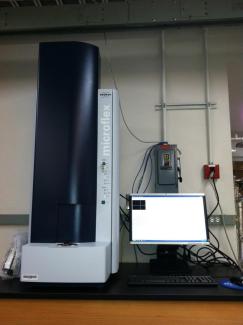
MALDI-TOF Tandem Mass Spectrometer
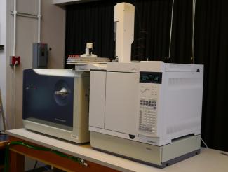
GCT Premier TOF Mass Spectrometer
Nuclear Magnetic Resonance (NMR) Facility
NMR spectroscopy is a highly sensitive technique in probing molecular structure and dynamics of chemical compounds, polymers and biomolecules. The in-house NMR Facility is currently home to five solution-state NMR spectrometers at 400, 500, and 600 MHz fields. Among them is a newly installed, state-of-the-art 500 MHz Bruker spectrometer equipped with a cryoprobe and an automatic sample lift. All spectrometers are equipped with multiple solution-state NMR probes for direct or indirect detection of 1H/19F and low frequency nuclei such as 13C, 31P, and 15N. These spectrometers are dedicated to the needs of chemistry and materials research with the 600 MHz instrument also used for biomolecular NMR research. Experiments conducted at the Facility range from routine 1D and 2D experiments for small molecule and polymer analysis at natural abundance to complex 3D and 4D multinuclear correlated experiments for proteins using isotope-labeled samples. All instruments are user-operated and are open to researchers with 24/7 access after proper training. User training is offered throughout the year in mostly hands-on individual sessions. Technical assistance is readily available for data collection and analysis.
The NMR lab of the Department of Chemistry and Biochemistry collaborates with the Spectroscopy Facility of the Materials Research Laboratory (MRL). The MRL-supported NMR facility is also available to all users on campus, and offers complementary capabilities, including solid state NMR instrumentation from 400 to 800 MHz, a CW Electron Paramagnetic Resonance (EPR) spectrometer and NMR imaging and diffusometry instrumentation.
Facility Manager and Spectroscopist: Dr. Hongjun Zhou; hzhou@chem.ucsb.edu
Location: Physical Sciences Building North (PSBN), Room 3614A
Phone: 805-893-2938
Wepage: NMR Facility
Dr. Hongjun Zhou
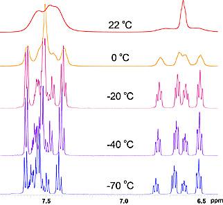
Changes in 1H spectrum in a variable temperature (VT) experiment indicates exchange among multiple conformers of a chemical compound
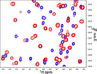
2D 1H-15N correlation spectrum shows perturbations to the protein amide resonances upon DNA binding
Optical Characterization Facility
In the Optical Characterization Facility, light is used to probe the intrinsic properties of materials prepared by chemists and serves as an analytical and quality control tool. We study energy and charge migration in materials for novel optoelectronic devices, test new biosensors and materials for data storage, zap bacteria with laser beams, and provide state of the art technical capabilities that are not commonly available to non-expert chemists in their own laboratory. The optical characterization facility gives students the ability to have hands-on experience in modern optical spectroscopy. The facility does not have dedicated instruments; rather experiments are designed to meet the need of the user. The facility manager assists its users in the design, assembly, and final analysis of experimental data. Custom optical experiments and support include photoconductive atomic force microscope, solar simulators for photovoltaics research or optical setups for characterization of organic semiconductors and many more chemical, materials and biological systems.
Major Capabilities and Areas of Expertise:
Time-resolved fluorescence measurements in the range 50 ps – 10 s (time-correlated single-photon counting etc.).
Transient absorption spectroscopy with sub-100 fs resolution
Custom luminescence spectroscopy (temperature-dependence, photoluminescence up-conversion, thermoluminescnce, electroluminescence, quantum-yield measurements)
Non-linear optical spectroscopy (multiphoton absorption and optical harmonics spectroscopy)
Raman spectroscopy and microscopy
High laser power processing of materials
Optical holography
Facility Instrumentation include 1 kHz repetition rate, 100 fs pulse Ti:Sapphire amplifier, Ti:Sapphire femtosecond oscillator laser, optical harmonics generator, high power Ar-ion laser with single-mode operation capability, various smaller semiconductor and gas lasers. We also have several monochromators equipped with spectroscopic CCD cameras, single channel detectors and a high-end Raman microscope.
Facility Manager and Spectroscopist: Dr. Alexander Mikhailovsky; mikhailovsky@chem.ucsb.edu
Location: Chemistry 3312
Phone: 805-893-2327
Webpage: Optical Characterization Facility
Dr. Alexander Mikhailovsky
X-Ray Facility
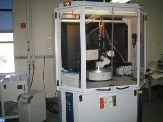
The in-house X-Ray facility allows single-crystal x-ray structure determination for researchers in the Department of Chemistry and Biochemistry at UCSB, as well as campus-wide. There are two main instruments currently housed in the facility. The micro-focus Mo and Cu duo source with direct photon count detector is used for chemical and protein crystallography and 2-D powder x-ray diffraction. Another diffractometer with a conventional sealed Mo x-ray tube is dedicated to small molecule structure determination. The following two x-ray instrumentations are available:
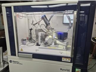
The Rigaku Synergy Diffractometer includes Rigaku Synergy XtaLab with a photon count detector and duo micro-focus X-ray sources. It has very bright focused beams of 0.1mm diameter area at sample site, and can be switched between Mo and Cu X-ray sources seamlessly in 1 min, and can collect data from crystals as small as 10 micron. The photon count detector HyPix-Arc 100, with the latest technology to detect X-ray photon directly, has little background noise, enabling decent data with long exposure if needed. The curved detector screen can cover as wide as 100 degree of 2theta area per frame, making data collection much more efficient. The system is equipped with an Oxford 1000 plus liquid nitrogen cryostream. The data collection can be operated between 80K and 500K, allowing researchers to work on air/moisture sensitive samples, explore the temperature dependent phase transition and other investigation. With Cu X-ray source, it is also able to run screening of protein crystals and determine absolute structure without presence of heavy elements. The 4-axis diffractometer allows identification of crystal faces, oriented X-ray scan and other analyses. With the adjustable beam slits, the fine focused intense Mo and Cu X-ray can collecting high resolution 2D powder diffractions, providing an option to run speedy PXRD with a little amount of sample and multi X-ray sources.
The Bruker Kappa APEX II Single-Crystal Diffractometer has a fine focus 2Kw Mo sealed tube x-ray source with a CCD detector and TRIUMPH monochromator. It is also equipped with a Bruker 4 axis diffractometer, an APEX II detector, Shelx software, and an Oxford 700 Plus Cryostream low temperature device — which can perform temperature-dependent measurement from 80K to 500K.
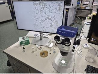
The Zeiss Stemi 305 Microscope performs crystal screening using a Polarized Lens, High Resolution Digital Camera and monitors. With an optical zoom up to 64x, the high resolution image display on the monitor can help pick the crystal(s), especially useful for demo and student training.
Faculty and students also have access to the complementary MRL X-ray facility, located just a few steps from the Chemistry building.
Facility Manager and Crystallographer: Dr. Guang Wu; wu@chem.ucsb.edu
Location: Physical Sciences Building North (PSBN), Room 4608
Phone: 805-893-2399
Webpage: X-ray Facility
Recharge Rate:
- Low Temperature Single Crystal Data collection: $200.00/sample
- Low Temperature Single Crystal Data collection and Structure Determination: $252.00/sample
- Room Temperature Single Crystal Data collection: $150.00/sample
- Room Temperature Single Crystal Data collection and Structure Determination: $202.00/sample
- Single Crystal Cell Determination:$30.00/sample
- Other Special Service: Rate depending on the nature of the experiment and measurement time